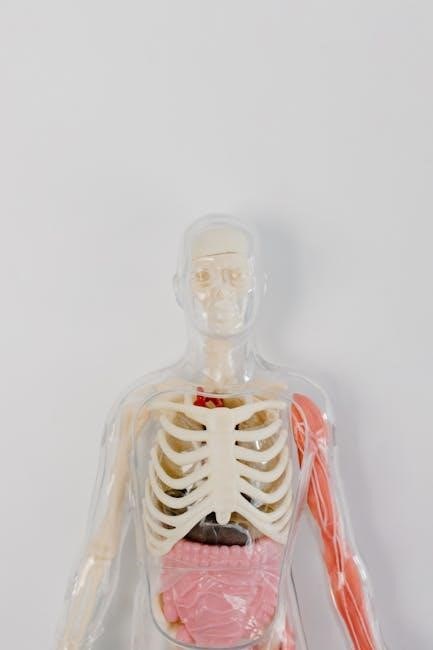
The humerus, the largest bone in the upper limb, connects the shoulder and elbow joints, providing structural support and attachment points for muscles and ligaments essential for upper limb mobility and movement.
1.1 Overview of the Humerus Bone
The humerus is the longest and thickest bone in the upper limb, extending from the shoulder to the elbow. It is a long bone with a cylindrical shaft and two expanded ends. The proximal end forms the head, which articulates with the glenoid cavity of the scapula, while the distal end includes the condyles that connect to the forearm bones. The bone features prominent landmarks such as the greater and lesser tubercles near the shoulder and the medial and lateral epicondyles near the elbow. These structures serve as attachment points for muscles and ligaments, enabling complex movements of the arm and forearm;
1.2 Importance of Humerus in Upper Limb Function
The humerus is crucial for upper limb function, serving as the structural foundation for arm movement and stability. It connects the shoulder and elbow joints, facilitating a wide range of motions such as flexion, extension, and rotation. Muscles like the deltoid, biceps, and triceps attach to its surface, enabling actions like lifting, throwing, and gripping. Additionally, the humerus protects vital nerves and blood vessels, such as the radial nerve, which runs through the spiral groove. Its durability and anatomical design allow for both strength and flexibility, making it indispensable for daily activities and complex motor functions. Damage to the humerus can significantly impair upper limb mobility and overall quality of life.
Proximal Humerus Anatomy
The proximal humerus includes the head, anatomical neck, and tubercles, forming the glenohumeral joint and serving as attachment points for muscles, enabling shoulder mobility and stability.
2.1 Humerus Head and Glenohumeral Joint
The humerus head, a rounded structure at the proximal end, articulates with the glenoid cavity of the scapula, forming the glenohumeral joint. This joint allows for extensive shoulder mobility, including flexion, extension, abduction, and rotation. The humerus head is supported by the anatomical neck, a slight constriction below it, which serves as an attachment point for the joint capsule and ligaments. The glenohumeral joint is crucial for upper limb function, enabling activities like throwing and reaching. Its stability is enhanced by surrounding muscles and ligaments, though it remains prone to dislocations and fractures, particularly in older adults due to bone density loss.
2.2 Anatomical Neck and Tubercles
The anatomical neck of the humerus is a narrow region located just distal to the humerus head. It serves as the attachment point for the joint capsule of the glenohumeral joint and is surrounded by the supraspinatus, infraspinatus, and teres minor muscles. Distal to the anatomical neck are the greater and lesser tubercles, which are prominent bony projections. The greater tubercle is the larger of the two and is located laterally, while the lesser tubercle is smaller and situated anteriorly. These tubercles provide Insertion points for the rotator cuff muscles, which stabilize the shoulder and facilitate movements such as abduction, internal rotation, and external rotation. The area between the tubercles is known as the intertubercular groove, which houses the long head of the biceps brachii tendon.
2;3 Greater and Lesser Tubercles
The greater and lesser tubercles are bony prominences located on the proximal humerus, serving as attachment sites for muscles of the shoulder. The greater tubercle, located laterally, provides insertion points for the supraspinatus, infraspinatus, and teres minor muscles, which are part of the rotator cuff. The lesser tubercle, situated anteriorly, is the insertion point for the subscapularis muscle. Together, these muscles stabilize the glenohumeral joint and enable movements such as abduction, internal rotation, and external rotation of the arm. The area between the tubercles, known as the intertubercular groove, contains the long head of the biceps brachii tendon, which plays a role in elbow flexion and forearm supination. These structures are crucial for upper limb function and mobility.

Distal Humerus Anatomy
The distal humerus includes the condyles and epicondyles, facilitating elbow joint movement and providing attachment points for forearm muscles, essential for flexion, extension, and supination.
3.1 Condyles and Epicondyles
The distal humerus features two condyles, medial and lateral, which form the elbow joint, enabling flexion, extension, and rotation. The medial condyle articulates with the ulna, while the lateral condyle connects with the radius. Above these condyles are the epicondyles, bony prominences that serve as attachment points for forearm muscles. The medial epicondyle anchors flexor muscles, and the lateral epicondyle anchors extensor muscles. These structures are crucial for forearm mobility and stability, with the olecranon fossa and radial fossa located nearby to accommodate the olecranon and radial head during joint movement. The condyles and epicondyles are vital for the functional anatomy of the elbow, facilitating daily activities like lifting and gripping.
3.2 Articular Surfaces and Forearm Mobility
The distal humerus features articular surfaces that form the elbow joint, facilitating forearm mobility. The medial and lateral condyles articulate with the ulna and radius, enabling flexion, extension, and rotation. These surfaces are covered with hyaline cartilage, reducing friction during movement. The olecranon and coronoid processes of the ulna interact with the humerus, allowing a wide range of motion. The radial head articulates with the capitellum of the humerus, contributing to supination and pronation. These articular surfaces are crucial for the functional anatomy of the elbow, enabling activities like lifting, gripping, and rotational movements. Their integrity is essential for maintaining forearm mobility and overall upper limb function. Damage to these surfaces can significantly impair movement and dexterity.

Muscle Attachments and Function
The humerus serves as a critical attachment point for muscles of the upper limb, enabling movements like flexion, extension, and rotation. Proximal muscles, such as the deltoid, supraspinatus, and infraspinatus, facilitate shoulder movements, while distal muscles, including the biceps brachii and triceps brachii, control elbow flexion and extension. These attachments ensure efficient biomechanical function, supporting daily activities and complex motor tasks.
4.1 Proximal Muscle Attachments
The proximal humerus is the attachment site for several key muscles that facilitate shoulder movement. The deltoid muscle attaches to the deltoid tuberosity, enabling abduction and flexion of the arm. The supraspinatus, infraspinatus, and teres minor muscles originate from the greater tubercle, contributing to external rotation and stabilization of the glenohumeral joint. The subscapularis muscle attaches to the lesser tubercle, playing a role in internal rotation and joint stability. Additionally, the pectoralis major, latissimus dorsi, and teres major muscles anchor to the proximal humerus, aiding in adduction, extension, and medial rotation. These muscle attachments collectively ensure precise and dynamic movement of the upper limb.
4.2 Distal Muscle Attachments
The distal humerus serves as the attachment site for muscles primarily involved in forearm and wrist movements. The medial epicondyle provides a attachment point for the flexor muscles of the forearm, including the flexor carpi radialis, flexor digitorum superficialis, and palmaris longus. The lateral epicondyle is the origin for the extensor muscles, such as the extensor carpi radialis brevis and extensor digitorum. Additionally, the brachialis muscle attaches near the distal humerus, contributing to elbow flexion, while the brachioradialis and triceps brachii also have attachment points in this region. These distal muscle attachments are crucial for controlling wrist and finger movements, enabling precise hand function and dexterity.
Blood Supply and Innervation
The humerus receives its blood supply primarily from the axillary and brachial arteries, with nutrient arteries penetrating the bone to maintain vitality. Innervation is provided by the radial and ulnar nerves.
5.1 Arterial Supply to the Humerus
The humerus receives its arterial supply primarily from the axillary artery and its continuation, the brachial artery. The axillary artery gives off branches, including the anterior and posterior circumflex humeral arteries, which supply the proximal regions and the humeral head. The brachial artery, running along the medial aspect of the humerus, provides the majority of blood flow to the bone, with nutrient arteries penetrating the cortex to supply the medullary cavity. Additional supply comes from the profunda brachii artery, which contributes to the vascular network around the bone. This extensive network ensures adequate perfusion, essential for bone health and function.
5.2 Nerve Distribution and Sensory Function
The humerus is closely associated with key nerves of the upper limb, including the radial, ulnar, and median nerves. The radial nerve runs in the spiral groove along the posterior aspect of the humerus, supplying the triceps brachii and extensor muscles of the forearm. The ulnar nerve passes near the medial epicondyle, providing sensation to the little finger and half of the ring finger. The median nerve, while not directly attached to the humerus, courses through the arm and forearm, innervating flexor muscles. Sensory innervation around the humerus is provided by cutaneous branches of these nerves, ensuring sensation over the arm and forearm. This complex nerve distribution is vital for motor function and sensory perception in the upper limb.

Fractures of the Humerus
Humerus fractures are classified based on location, with proximal fractures near the shoulder and distal fractures near the elbow, often requiring surgical intervention for proper alignment and healing.
6.1 Classification of Humerus Fractures
Humerus fractures are classified based on their location and characteristics. Proximal fractures occur near the shoulder, while distal fractures are closer to the elbow. The Neer classification system is commonly used for proximal humerus fractures, categorizing them into two main types: one-piece fractures (non-displaced) and multi-fragment fractures. Distal humerus fractures are often classified using the AO/OTA system, which groups them into extra-articular (A), partial articular (B), and complete articular (C) fractures. The classification considers factors such as displacement, comminution, and involvement of the articular surface. Accurate classification is essential for determining the appropriate treatment and predicting outcomes. Each type of fracture requires a tailored approach to ensure proper alignment and functional recovery.
6.2 Treatment Options for Proximal and Distal Fractures
Treatment of humerus fractures varies based on the fracture’s location and severity. For proximal fractures, non-surgical options like immobilization may be used for non-displaced fractures, while displaced fractures often require surgical intervention, such as open reduction and internal fixation (ORIF) or hemiarthroplasty. Distal fractures frequently involve articular surfaces, necessitating precise alignment to restore joint function. Internal fixation with plates or screws is common, especially for unstable or displaced fractures. In some cases, anatomical locking plates are used to stabilize complex fractures. The choice of treatment depends on factors such as fracture pattern, patient health, and lifestyle. The goal is to restore anatomical alignment, achieve bone healing, and maximize functional recovery, particularly for the shoulder and elbow joints.
Clinical Relevance and Surgical Anatomy
The humerus’s complex anatomy is crucial in clinical settings, particularly for surgical interventions like osteotomies and arthroplasty, requiring precise understanding of its structural and functional components.
7.1 Surgical Approaches to the Humerus
Surgical approaches to the humerus vary based on fracture location and type. Proximal humerus fractures often require a deltopectoral approach, providing access to the glenohumeral joint and tubercles. Distal fractures may use a posterior approach, minimizing soft tissue damage. Internal fixation with anatomical locking plates is common for both proximal and distal fractures, ensuring stability and promoting healing. Open reduction is frequently necessary for articular shear injuries. Minimally invasive techniques are increasingly used to reduce complications and improve recovery times. Understanding the humerus’s surgical anatomy is critical for successful outcomes, emphasizing precise knowledge of muscle attachments and nerve distribution to avoid iatrogenic injury.
7.2 Humerus Osteotomies and Arthroplasty
Humerus osteotomies are surgical procedures involving precise bone cuts to correct deformities or improve alignment. These are often performed to address malunions or nonunions, ensuring proper healing and function. Arthroplasty, such as total shoulder or elbow replacement, is used when joint surfaces are severely damaged. Proximal humerus arthroplasty is common for fractures or arthritis, while distal arthroplasty addresses elbow joint degeneration. Surgical techniques emphasize preserving bone stock and soft tissue integrity. Advances in implant design, including anatomical shoulder prostheses, aim to restore mobility and reduce complications. These procedures require meticulous planning and execution, highlighting the importance of understanding humeral anatomy for optimal outcomes and patient recovery.

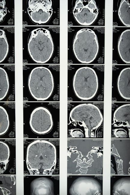
Imaging Techniques for Humerus Anatomy
Imaging techniques like X-rays, MRIs, and CT scans are essential for evaluating humerus anatomy, providing detailed views of fractures, joint alignment, and soft tissue integrity for accurate diagnoses and treatment planning.
8.1 Radiographic Evaluation of the Humerus
Radiographic evaluation of the humerus is critical for assessing its structure and identifying injuries or abnormalities. Common imaging techniques include X-rays, CT scans, and MRIs. X-rays are often the first-line imaging method, providing clear views of fractures, dislocations, and bone alignment. CT scans offer detailed cross-sectional images, particularly useful for complex fractures or articular surface injuries. MRIs are ideal for evaluating soft tissue injuries, such as tendon or ligament tears, and for detecting subtle fractures not visible on X-rays. Proper positioning and multiple views are essential to ensure accurate diagnosis. These imaging modalities are indispensable for diagnosing humerus-related conditions, guiding treatment plans, and monitoring recovery in both acute and chronic cases.

8.2 Advanced Imaging Modalities
Advanced imaging modalities, such as MRI and CT scans, are crucial for detailed evaluation of the humerus. MRI provides exceptional visualization of soft tissues, including muscles, tendons, and ligaments, aiding in the diagnosis of injuries like tendon tears or ligament sprains. CT scans offer high-resolution images of bone structures, making them ideal for assessing complex fractures, articular surfaces, and bone density. These modalities complement radiographic evaluation by providing deeper insights into both bone and soft tissue abnormalities. They are essential for preoperative planning, monitoring recovery, and ensuring accurate diagnoses in cases where X-rays are insufficient. Advanced imaging enhances the ability to detect and manage a wide range of humerus-related conditions effectively.
The humerus, as the longest bone in the upper limb, plays a pivotal role in movement and structural support. Its complex anatomy, including the proximal and distal regions, muscle attachments, and articular surfaces, underscores its importance in upper limb function. Understanding the humerus’s anatomy is crucial for diagnosing and treating fractures, injuries, and surgical interventions. Advanced imaging techniques and surgical approaches highlight the bone’s clinical relevance. This comprehensive overview emphasizes the humerus’s vital role in mobility and its intricate relationship with surrounding tissues. Continued study of its anatomy remains essential for advancing both clinical and surgical practices, ensuring improved outcomes for patients with humerus-related conditions.